Companion animal guardians frequently encounter escalating behavioral resistance and severe skin complications when attempting routine coat maintenance and claw trimming in home environments. Beneath seemingly superficial aesthetic tangles, unmanaged shedding undercoats rapidly form dense felted mats that trap moisture against the epidermal stratum corneum, creating hypoxic incubators for severe microbial dermatitis, painful cutaneous ulcerations, and crippling claw deformities. Attempting to forcibly restrain a panic-stricken pet with unsuitable human shears or aggressive pulling transforms basic hygiene into traumatic encounters that induce severe veterinary handling phobias and accidental lacerations. By implementing evidence-based low-stress desensitization protocols, anatomical line brushing techniques, and pH-balanced dermatological grooming strategies, caregivers preserve cutaneous barrier homeostasis while establishing reliable cooperative care partnerships that ensure lifelong physiological wellbeing.
1. Cutaneous Histology and Hair Follicle Biology Across Companion Animal Breeds
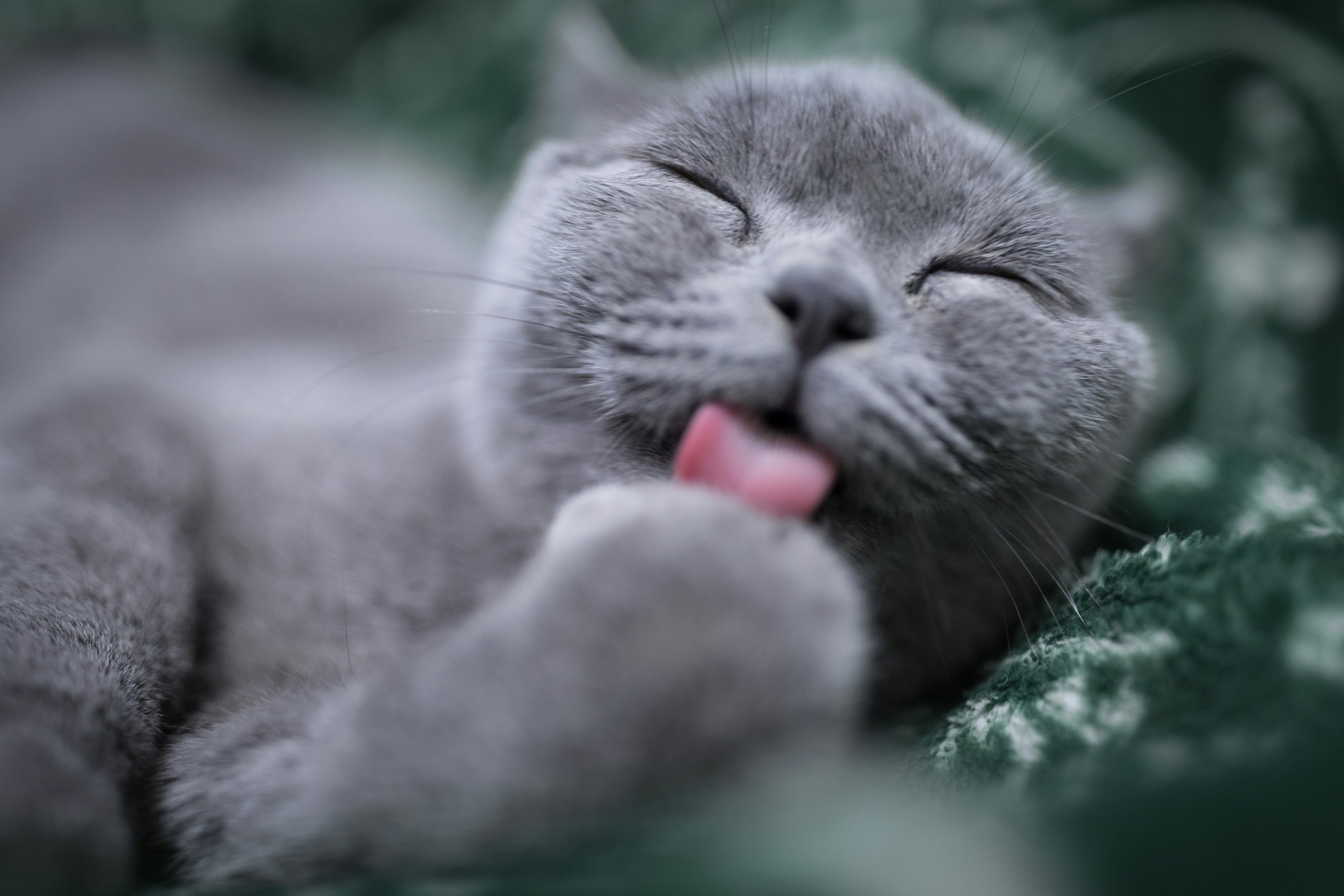
Low-stress desensitization and gradual tactile habituation allow companion animals to perceive coat grooming as a predictable, reinforcing welfare protocol. Developing an effective home grooming protocol requires an understanding of mammalian integumentary anatomy. Canine and feline skin differs fundamentally from human skin in epidermal thickness, cellular turnover, and chemical buffering capacity. While human epidermis comprises ten to fifteen nucleated cellular layers, canine and feline epidermis measures merely three to five cellular strata in non-follicular regions, rendering companion animal skin exceptionally fragile and prone to mechanical abrasion during brushing.
Furthermore, companion animals possess compound hair follicle arrangements where multiple fine secondary undercoat hairs emerge from a single follicular ostium alongside a central primary guard hair. Understanding the functional classification of coat types governs instrument selection and mechanical technique:
- Short Smooth Coats: Breeds such as Pugs, Boxers, and Domestic Shorthair cats feature dense primary hairs with minimal undercoat. Natural epidermal sebum distribution is best stimulated using flexible rubber curry brushes that exfoliate dead keratinocytes without scratching dermal papillae.
- Double Coats: Pastoral, spitz, and retriever breeds (including German Shepherds, Siberian Huskies, and Golden Retrievers) possess harsh water-repellent guard hairs supported by dense, woolly undercoats designed for dual-phase thermal regulation. Seasonal follicular shedding cycles require specialized undercoat rakes to prevent felting.
- Continuous-Growth Non-Shedding Coats: Poodles, Bichon Frises, and related hybrid breeds feature fine, curly keratin filaments that lack an active seasonal telogen shedding drop. Shed hairs become mechanically entrapped within surrounding curls, necessitating daily line brushing down to the dermal margin to prevent cast-like matting.
- Wire Coats: Terriers feature coarse guard hairs with tapered tips that require manual hand-stripping or specialized undercoat clearing to maintain follicular cycle turnover and natural water-resistant texture.
A critical veterinary dermatological imperative involves the strict prohibition against shaving double-coated companion animals. Shaving removes protective primary guard hairs that reflect solar radiation and shield delicate skin from ultraviolet burns. Post-clipping alopecia frequently ensues, characterized by prolonged follicular arrest where undercoat regrows erratically while guard hairs remain dormant, permanently compromising thermoregulatory efficiency and predisposing the animal to solar dermatitis.
2. Specialized Instrumentation and Mechanical Line Brushing Methodologies
Attempting to detangle a companion animal’s coat using superficial surface brushing merely grooms the upper layer of guard hairs while leaving dense undercoat mats to tighten silently against the skin. Systematic line brushing represents the gold-standard clinical technique for complete undercoat management.
Line brushing begins by parting the hair with the non-dominant hand until the skin is clearly visible along a linear boundary. Utilizing an ergonomic slicker brush with polished, flexible stainless-steel pins, the handler brushes downward in short, gentle pat-and-pull strokes along the parted line. Once a section moves smoothly, the parted boundary is shifted incrementally across the torso. Every completed section is subsequently validated using a stainless-steel greyhound comb, whose long tines verify that no subterranean knots remain adjacent to the dermis.
| Coat Category | Representative Breeds | Primary Instrumentation | Clinical Technique & Preventative Frequency |
|---|---|---|---|
| Double Coat | Golden Retriever, Siberian Husky, German Shepherd | Rotating undercoat rake, Long-pin slicker brush | Sectional line brushing 3x weekly; high-velocity cool air drying during seasonal coat blow |
| Curly / Non-Shedding | Poodle, Bichon Frise, Doodle cross-hybrids | Flexible slicker brush, Stainless-steel greyhound comb | Daily line brushing down to the dermal margin; detangling hydration spray to reduce pin friction |
| Short / Smooth | Beagle, Doberman Pinscher, DSH feline | Natural bristle brush, Flexible rubber curry pad | Circular currying weekly to dislodge telogen shafts, exfoliate, and distribute lipid sebum |
| Long / Silky | Yorkshire Terrier, Maltese, Persian cat | Pin brush (no protective balls), Wide-tooth metal comb | Daily gentle stroke detangling; focus on high-friction axillary (armpit) and inguinal zones |
Clinical Surgical Warning — Severe Matting, Vascular Strangulation, and Reperfusion Pinna Hematomas: Beyond cosmetic and dermatological compromise, dense felted matting presents severe vascular and surgical hazards. Compacted hair casts exert unrelenting mechanical tension on underlying dermal margins, strangling local capillary microcirculation (vascular strangulation) for weeks or months. This vascular compromise becomes exceptionally dangerous along the ear leather (pinna) and tail tip. When dense mats are clipped away, the sudden relief of external compression triggers a dramatic reperfusion surge as arterial blood abruptly engorges previously constricted, fragile capillary beds. This sudden hemodynamic rush frequently ruptures weakened vessels, causing massive aural hematomas (pinna hematomas) or relentless margin hemorrhage as the animal violently shakes its head in response to localized tingling paresthesia. Veterinary surgical and professional grooming standards mandate that immediately following the removal of severe ear matting, handlers must apply a protective compressive head wrap, snood, or elastic calming band (such as a Happy Hoodie) for 24 to 48 hours to restrict vigorous head shaking and prevent catastrophic hematoma formation.
3. Digital Claw Biomechanics, Vascular Quick Anatomy, and Cooperative Trimming
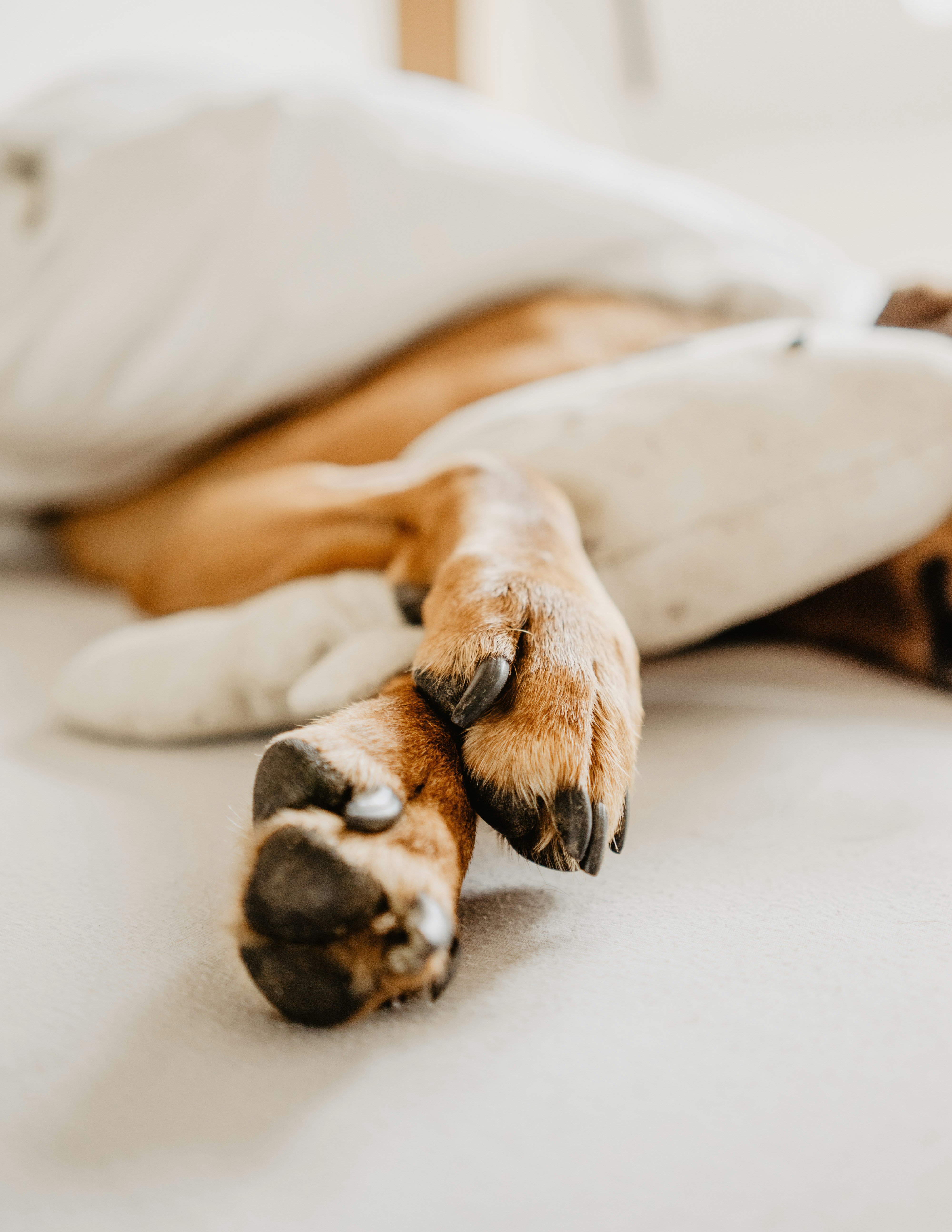
Precise claw shortening prevents metacarpophalangeal hyperextension while avoiding laceration of the vascular quick. Canine and feline claws represent modified keratinous integumentary structures firmly anchored to the third phalanx (P3) of each digit. Within each claw lies the dermis unguis, colloquially termed the vascular quick, which contains high-density sensory nerve endings and an abundant capillary network that supplies the germinal ungual matrix.
When claw trimming is neglected, elongated nails disrupt normal posture. During standing and ambulation, excessively long claws strike the floor prematurely, forcing the third phalanx into continuous retroflexion. This mechanical distortion alters ground reaction force vectors through the digital flexor tendons, causing compensatory hyperextension of the metacarpophalangeal joints, chronic postural strain, and accelerated osteoarthritis across the carpal, cubital, and coxofemoral joints.
Trimming requires precise anatomical visualization. In unpigmented nails, the pink vascular cone is readily identifiable against the translucent dorsal claw wall. The incision should be angled parallel to the natural slope of the paw pad, terminating approximately two millimeters distal to the quick. In darkly pigmented claws where internal vascular borders are obscured, handlers must employ progressive micro-slices with sharp guillotine or bypass clippers, or utilize a rotary diamond drum grinder. As slicing progresses, the emergence of a homogenous grey or black oval core indicates proximity to the apical quick boundary, signaling that further reduction should cease immediately.
| Conditioning Phase | Target Sensory Stimulus | Reinforcement Mechanism & Pairing | Advancement Criteria to Next Phase |
|---|---|---|---|
| Phase 1: Tactile Habituation | Gentle digital paw squeeze and individual toe separation | High-value primary reward delivered immediately upon touch release | Zero paw withdrawal reflex across 10 consecutive clinical trials |
| Phase 2: Instrument Introduction | Metal clipper / grinder tactile touch against outer claw wall | High-value lick mat pairing (peanut butter or wet food paste) | Calm autonomic tone; soft eye gaze and relaxed body posture maintained |
| Phase 3: Acoustic Desensitization | Rotary grinder motor hum or dry pasta clipping sound nearby | Continuous secondary reinforcer delivery during active sound emission | Absence of ear pinning, lip licking, yawning, or postural freezing |
| Phase 4: Operant Micro-Trimming | Single distal claw sliver reduction (1mm) per training session | Immediate jackpot food reward delivered upon single micro-cut | Voluntary chin rest or paw presentation maintained without restraint |
Clinical Hemostatic Fact — Ferric Subsulfate Chemical Burns and the Non-Rubbing Pinpoint Pressure Rule: Accidental laceration of the vascular quick requires immediate, evidence-based hemostatic intervention to prevent both hemorrhage and behavioral trauma. Veterinary styptic powders contain ferric subsulfate (or ferric chloride), an aggressive chemical protein coagulant that forms an instantaneous micro-thrombus. However, a widespread and destructive caregiver error is vigorously rubbing or grinding styptic powder into the bleeding claw surface. Rubbing forces caustic chemical salts directly into exposed sensory nerve endings, causing excruciating chemical burning pain that instills severe handling phobias and subsequent defensive aggression. The correct clinical protocol mandates pinpoint pressure (the non-rubbing pinpoint rule): handlers must apply a small quantity of styptic powder using a moistened cotton swab or fingertip pressed firmly and perpendicularly against the bleeding apical claw surface for 5 to 10 seconds without lateral friction. In domestic environments where commercial styptic powder is unavailable, pure non-irritating cornstarch or flour provides a safe, sting-free mechanical tamponade that promotes clotting without chemical tissue irritation.
4. Epidermal Acid Mantle Preservation and Bathing Chemistry
Maintaining epidermal lipid integrity requires species-specific, pH-buffered surfactants formulated to preserve the canine acid mantle. One of the most pervasive home grooming errors is the application of human cosmetic shampoos or dishwashing detergents to domestic pets. Mammalian epidermal surface pH differs dramatically across phylogenetic families. While normal human skin maintains an acidic buffering mantle ranging between pH 5.2 and 5.8, canine skin exhibits a near-neutral to mildly alkaline pH spectrum between 6.5 and 7.5. Feline epidermal pH typically measures between 6.0 and 6.8.
Exposing companion animal skin to acidic human formulations destroys the delicate stratum corneum barrier. Acidic surfactants strip ceramides, free fatty acids, and cholesterol esters from the intercellular lipid matrix. This chemical disruption provokes trans-epidermal water loss (TEWL), leaving the skin severely desiccated and pruritic. Consequently, commensal bacterial flora (such as Staphylococcus pseudintermedius) and fungal yeasts (such as Malassezia pachydermatis) proliferate uncontrollably, triggering secondary superficial pyoderma and recurrent pruritus.
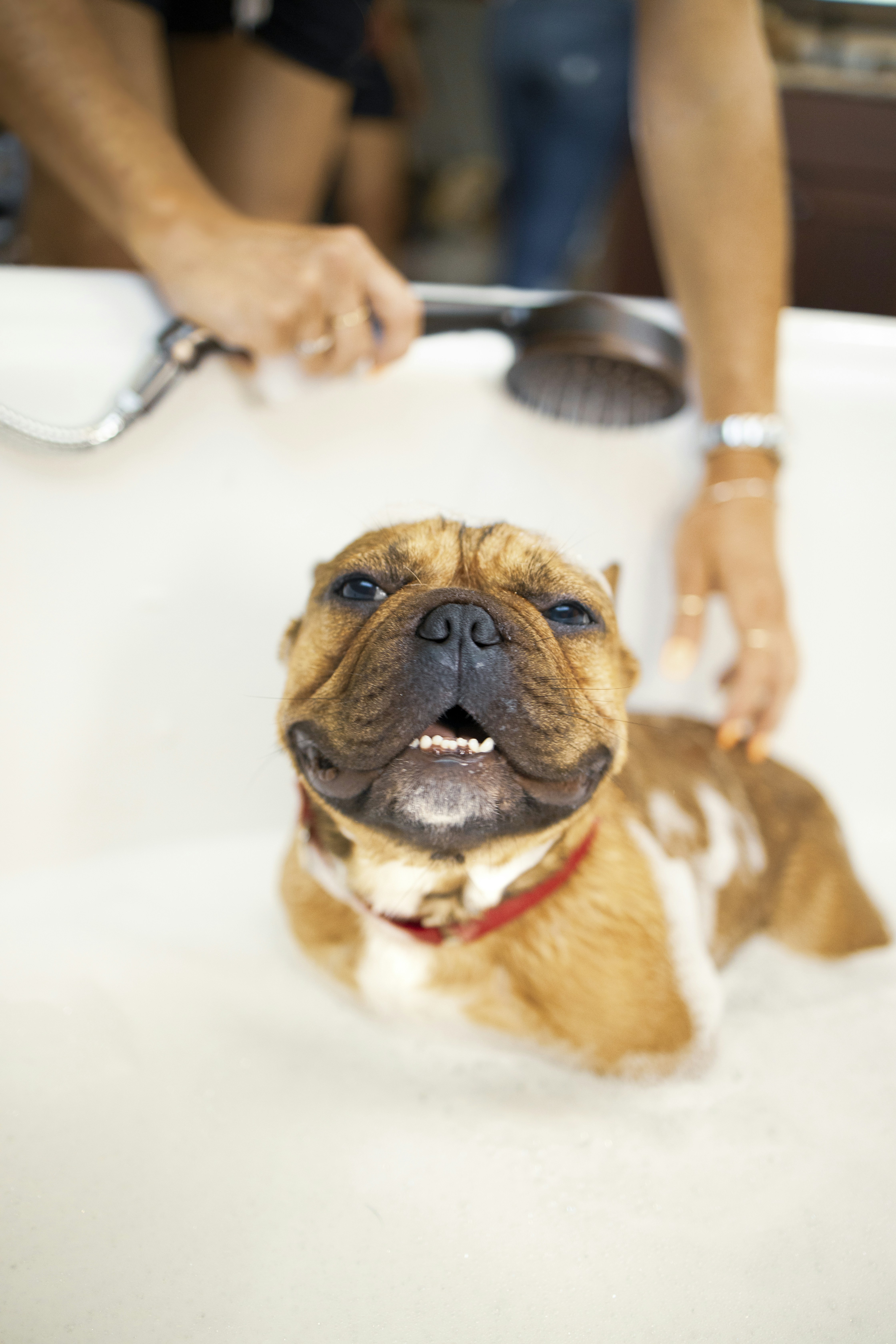
Home bathing regimens must exclusively utilize veterinary-formulated, pH-balanced products featuring gentle non-ionic or amphoteric surfactants alongside humectants (such as colloidal oatmeal, glycerin, and phytosphingosine). Bathing water temperature must remain tepid, as elevated water temperatures induce local cutaneous vasodilation, exacerbating underlying pruritus and triggering thermal panic. Drying must prioritize high-absorbency microfiber towels followed by cold-to-lukewarm velocity airflow, ensuring that heated air dryers never contact dermal tissue.
Veterinary Dermatological & Biomechanical Home Grooming Simulator
Quantify trans-epidermal water loss (TEWL), third phalanx (P3) postural strain, and autonomic panic indices across companion animal grooming regimens.
Critical Compromise
Active Epidermal Stripping & Articular Strain in Progress
85%
85%
65%
5. Stress-Free Cooperative Care Conditioning and Body Language Triage
Traditional coercive restraint—such as forcefully pinning an animal to a table or holding their muzzle shut during grooming—triggers sympathetic autonomic activation. When an animal experiences intense fear without the possibility of flight, acute defensive aggression frequently manifests as a desperate survival mechanism.
Modern veterinary behavioral science advocates for cooperative care protocols founded on choice, predictability, and positive reinforcement. Caregivers establish a designated “start button” behavior—such as the animal voluntarily placing their chin upon a cushioned towel or offering a paw onto a handler’s palm. Grooming actions (brush strokes, ear cleaning, or claw grinding) proceed only while the animal maintains this voluntary posture. The moment the animal lifts their chin or breaks contact, all grooming ceases immediately, reassuring the pet that they retain agency over physical manipulation.
During every grooming session, handlers must actively monitor somatic stress indicators. Lip licking, yawning outside of rest periods, brow furrowing, cutaneous twitching, and whale-eye (scleral exposure) signify escalating autonomic distress. Observing any of these subtle warning cues requires an immediate pause, lowering the intensity of manipulation, and pairing the session with high-value food reinforcers before fear conditioning solidifies.