Companion animal caregivers frequently dismiss early mobility decline in aging dogs and cats as inevitable chronological slowing, allowing progressive joint degradation to advance untreated. Because domestic pets retain deeply rooted evolutionary instincts to conceal physical discomfort from perceived predators and social rivals, insidious articular inflammation, subchondral micro-fractures, and compensatory spinal strain progress through protracted subclinical stages before overt lameness or vocalization emerges. By the time an animal displays visible limping or complete refusal to climb stairs, articular hyaline cartilage is severely denuded, locking the senior companion into agonizing cycles of chronic central pain sensitization and muscle atrophy. By implementing comprehensive veterinary multimodal pain management frameworks that combine targeted cyclooxygenase-2 inhibitors, novel monoclonal antibodies, nutritional chondroprotection, and proactive domestic biomechanical modifications, handlers arrest degenerative cascades and dramatically preserve lifelong locomotive vitality.
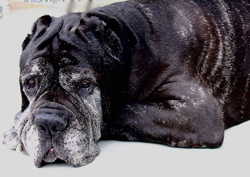
1. Osteoarthritis Pathophysiology and Chondrocyte Degradation Mechanisms
Osteoarthritis (OA), also termed degenerative joint disease (DJD), is a chronic, non-infectious, progressive disorder affecting diarthrodial synovial joints. Rather than a simple mechanical “wear and tear” phenomenon, OA is an active biochemical pathology driven by persistent low-grade synovial inflammation and catabolic cellular cascades within articular chondrocytes.
Under normal physiological conditions, articular hyaline cartilage provides a friction-free, shock-absorbing surface composed of an extracellular matrix rich in type II collagen and sulfated proteoglycans (primarily aggrecan). When mechanical instability—such as cranial cruciate ligament rupture, hip dysplasia, or chronic micro-trauma—disrupts biomechanical balance, chondrocytes release pro-inflammatory cytokines, predominantly interleukin-1 beta (IL-1β) and tumor necrosis factor-alpha (TNF-α). These cytokines up-regulate matrix metalloproteinases (MMPs) and aggrecanases, which enzymatically cleave structural collagen fibrils and deplete hydrophilic glycosaminoglycans.
As the extracellular matrix loses water retention capacity, cartilage fibrillates, develops vertical clefts, and gradually erodes to expose underlying subchondral bone. Compensatory periarticular osteophytosis (bone spur development) and subchondral sclerosis follow, generating continuous nociceptive firing from densely innervated subchondral vessels and inflamed synovial membranes.
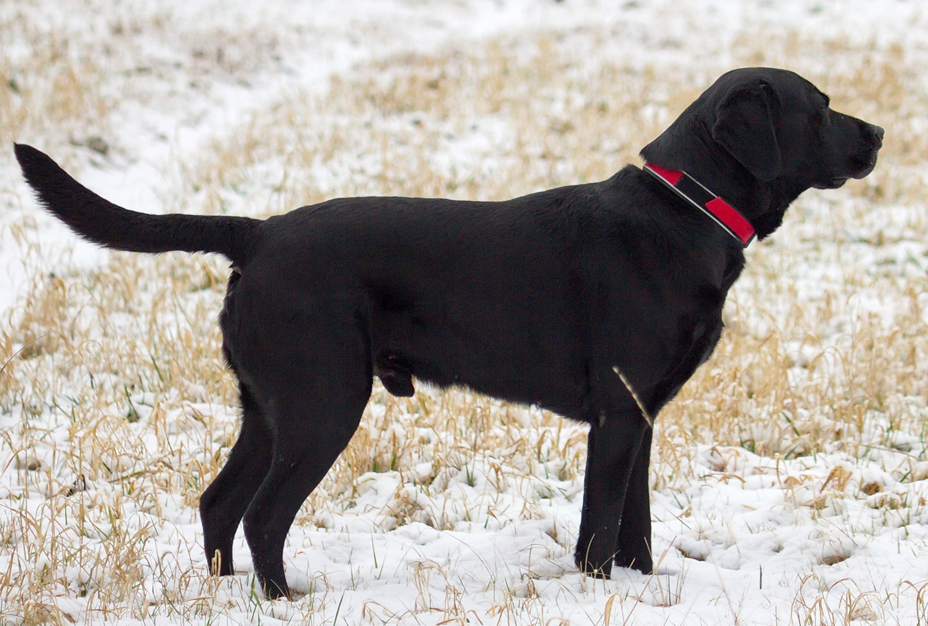
2. Species-Specific Clinical Symptomatology and the Feline Masking Trap
Canine and feline patients manifest orthopedic discomfort through divergent behavioral pathways. While dogs exhibit recognizable locomotive alterations, cats present subtle behavioral anomalies that owners frequently misinterpret as normal aging.
In canines, early clinical indicators include:
- Morning Stiffness & The Warm-Up Effect: Notable locomotive hesitation upon rising that temporarily improves after several minutes of ambulation.
- Hesitation on Vertical Inclines: Reluctance to jump into vehicles, ascend staircases, or leap onto furniture.
- Asymmetrical Muscular Atrophy: Bilateral differences in quadriceps, hamstring, or supraspinatus muscle mass secondary to chronic disuse and off-loading.
- Licking at Arthritic Joints: Repetitive licking over the carpi or stifies, occasionally causing secondary acral lick dermatitis.
Conversely, felines almost never exhibit overt limping unless affected by catastrophic trauma or end-stage single-limb pathology. Feline OA manifests predominantly as behavioral avoidance. Arthritic cats alter their trajectory by dividing jumps into intermediary steps (e.g., floor to low footstool, then to bed) rather than leaping directly. Furthermore, high-sided litter boxes require painful coxofemoral flexion to enter, causing senior cats to eliminate on flat floor surfaces immediately adjacent to the box. Unkempt, matted coats over the lumbosacral region and claw overgrowth secondary to reduced scratching-post use serve as definitive red flags for spinal and carpal arthritic pain.
| Clinical OA Stage | Functional Impairment | Primary Pharmacotherapy | Adjunctive Interventions |
|---|---|---|---|
| Stage 1: Subclinical / Mild | Intermittent stiffness after intense exercise | Marine Omega-3 (high-dose EPA/DHA), Green Lipped Mussel | Weight optimization (target BCS 4.5/9), low-impact leash walks |
| Stage 2: Moderate Chronic | Hesitation on stairs, altered gait, reduced grooming | COX-2 selective NSAIDs (Carprofen, Meloxicam, Robenacoxib) | Polysulfated glycosaminoglycan (Adequan) injections, home traction |
| Stage 3: Severe Progressive | Persistent lameness, muscle atrophy, reluctance to stand | Anti-NGF mAbs (Bedinvetmab / Librela, Frunevetmab / Solensia) | Gabapentin for central wind-up pain, class IV photobiomodulation |
| Stage 4: End-Stage Refractory | Severe recumbency, vocalization upon touch, central sensitization | Multimodal triple therapy: NSAID/mAb + Amantadine + Gabapentin | Palliative hydrotherapy, assisted mobility harnesses, acupuncture |
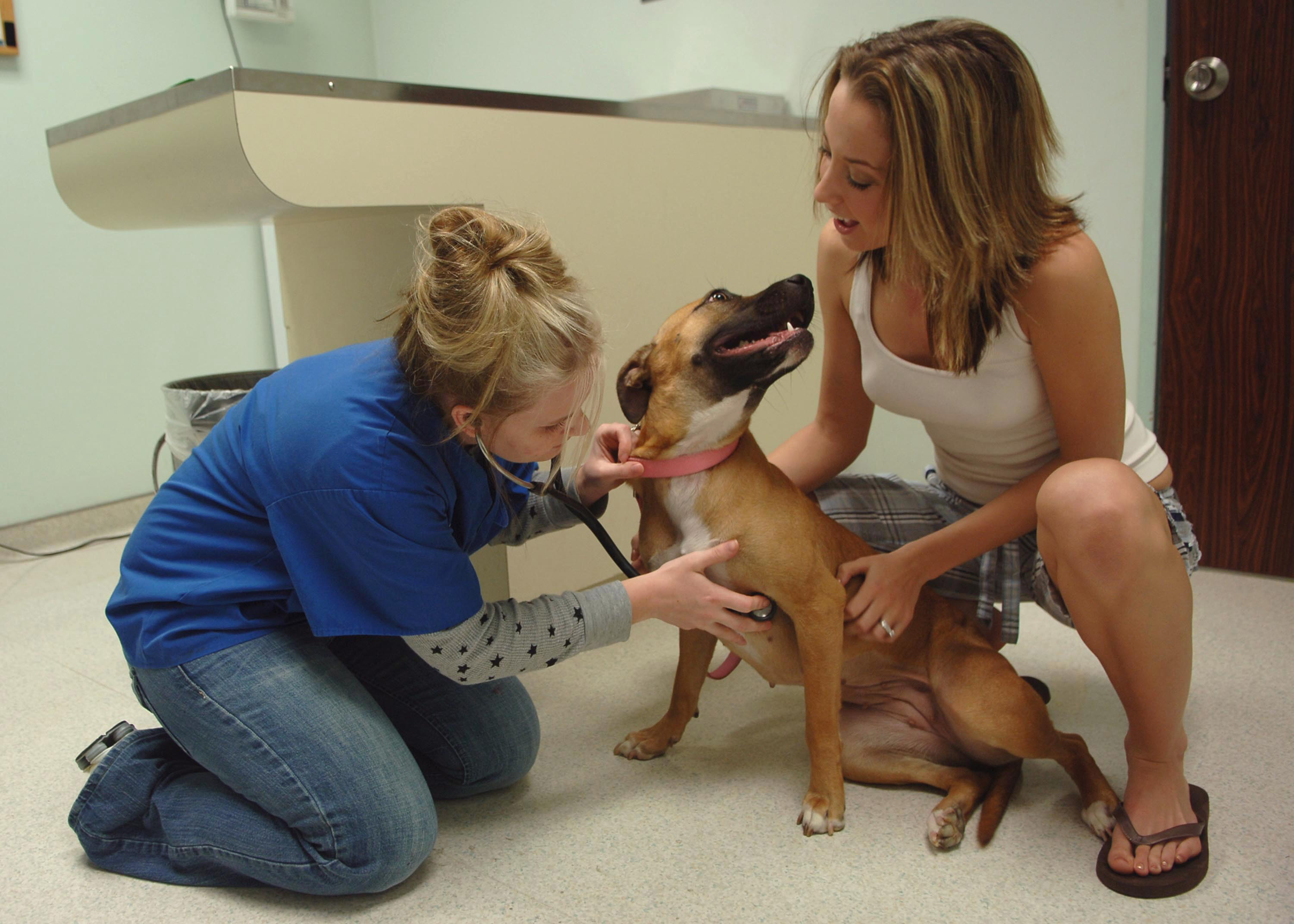
3. Multimodal Pharmacological Strategies: NSAIDs, Monoclonal Antibodies, and Neuropathic Agents
Modern veterinary pain management strictly rejects monotherapy in favor of the multimodal analgesic pyramid. Combining agents with distinct cellular mechanisms allows clinicians to disrupt nociceptive transmission at multiple neuro-anatomical levels while minimizing drug dosages and reducing adverse effects.
Non-Steroidal Anti-Inflammatory Drugs (NSAIDs) operating via selective cyclooxygenase-2 (COX-2) inhibition—such as carprofen, meloxicam, deracoxib, and robenacoxib—remain foundational first-line therapies. By inhibiting the enzymatic conversion of arachidonic acid into pro-inflammatory prostaglandin E2 (PGE2), NSAIDs alleviate peripheral nociceptor sensitization. However, pre-treatment screening and serial laboratory monitoring (CBC, renal chemistry, urinalysis, and hepatic panels) every six months are mandatory to safeguard against idiosyncratic nephrotoxicity or gastrointestinal mucosal ulceration.
The advent of species-specific anti-nerve growth factor (anti-NGF) monoclonal antibodies represents the most transformative milestone in companion animal orthopedics. Nerve growth factor is significantly elevated in arthritic synovial fluid, binding to tropomyosin receptor kinase A (TrkA) receptors on peripheral nociceptors and dorsal root ganglia to drive neuropathic hyperalgesia. Injectable monthly monoclonal antibodies—bedinvetmab (Librela) for dogs and frunevetmab (Solensia) for cats—specifically bind and neutralize circulating NGF. Because these therapeutic antibodies are metabolized through normal cellular protein catabolic pathways rather than hepatic cytochrome P450 or renal excretion, they provide safe, robust analgesia even in patients with concurrent early renal or hepatic comorbidities.
For patients suffering from chronic mal-adaptive pain, secondary neuropathic agents are indispensable. Oral gabapentin modulates voltage-gated calcium channels within dorsal horn neurons, suppressing spinal excitatory glutamate release. Concurrently, oral amantadine functions as an N-methyl-D-aspartate (NMDA) receptor antagonist, effectively attenuating central wind-up pain and reversing established hyperalgesia.
4. Nutritional Chondroprotection and Caloric Biomechanics
Biomechanical load management is the single most potent non-pharmacological determinant of arthritic disease progression. In overweight or obese companion animals, excess adiposity acts as a chronic endocrine organ that secretes systemic inflammatory adipokines (including leptin, adiponectin, and IL-6), fueling intra-articular inflammation. Furthermore, every additional kilogram of body mass magnifies joint reaction forces exponentially during locomotion.
Clinical studies demonstrate that a modest 6% to 8% body weight reduction significantly decreases lameness scores, reduces required NSAID dosages, and restores joint range of motion. Diets formulated for senior mobility must prioritize high protein-to-calorie ratios to preserve lean sarcopenic muscle mass while facilitating controlled caloric restriction.
Nutraceutical supplementation must rely on verified bioavailable compounds. Marine-derived long-chain omega-3 polyunsaturated fatty acids (specifically eicosapentaenoic acid, EPA, and docosahexaenoic acid, DHA) replace arachidonic acid within cell membrane phospholipid bilayers, shifting downstream eicosanoid synthesis toward anti-inflammatory series-3 prostaglandins and series-5 leukotrienes. Clinical veterinary nutrition guidelines recommend a therapeutic dosage of 100 to 150 mg combined EPA/DHA per kilogram of metabolic body weight. Conversely, traditional oral glucosamine and chondroitin sulfate supplements demonstrate highly erratic oral bioavailability and variable clinical efficacy unless paired with bio-enhanced formulations like undenatured type II collagen (UC-II) or green-lipped mussel extract.
| Living Environment Zone | Mechanical Hazard | Architectural Intervention | Biomechanical Outcome |
|---|---|---|---|
| Smooth Flooring | Hardwood and tile micro-slipping | Interlocking yoga mats, non-skid runners | Eliminates catastrophic groin adductor strains and falling panic |
| Vertical Elevation | High beds, sofas, vehicle trunks | Gradual incline carpeted ramps (slope ≤ 25°) | Removes high-impact compressive landing forces from carpi and elbows |
| Feline Elimination | Deep high-sided litter boxes | Low-entry boxes with 2-inch cutouts | Prevents coxofemoral hyperextension, resolving periuria accidents |
| Resting Substrates | Thin floor pads bottoming out | High-density orthopedic memory foam mattresses | Distributes body weight evenly, protecting bony prominences |
5. Physical Rehabilitation Modalities and Controlled Home Exercise
Complete cage rest is universally contraindicated in chronic osteoarthritis. Immobilization induces rapid periarticular ligamentous shortening, synovial fluid stagnation, and accelerated muscular atrophy, which exacerbates joint instability upon subsequent movement. Successful long-term management hinges upon consistent, low-impact physical exercise.
Canine exercise programs should prioritize multiple short, controlled leash walks (10 to 15 minutes, three times daily on level grass or packed dirt) rather than sporadic weekend excursions over rugged terrain. Incorporating controlled proprioceptive exercises—such as stepping over low obstacles (cavaletti rails), gentle weight-shifting exercises, and sit-to-stand transitions—maintains neuromuscular coordination and stabilizes supporting musculature.
Professional veterinary physical rehabilitation modalities significantly augment home management. Underwater treadmill hydrotherapy utilizes water buoyancy to offload up to 60% of gravitational body weight while providing hydrostatic pressure and thermal warmth that enhance joint mobility. Concurrently, Class IV therapeutic photobiomodulation (low-level laser therapy) stimulates mitochondrial cytochrome c oxidase, boosting ATP production, accelerating tissue repair, and dampening inflammatory pain signaling without systemic pharmacological burden.