Foul pet breath is frequently dismissed as an inevitable hallmark of animal aging, yet severe halitosis is a primary clinical indicator of active anaerobic bacterial proliferation destroying the periodontium. Left unaddressed, superficial marginal gingivitis silently progresses into irreversible periodontal ligament lysis, extensive alveolar bone resorption, and painful tooth loss. Even more alarming, continuous oral bacteremia seeds virulent pathogens directly into the systemic circulation, predisposing aging pets to vegetative valvular endocarditis, hepatic microabscesses, and chronic glomerulonephritis. A multi-modal dental prophylaxis strategy combining daily mechanical brushing, veterinary ultrasonic scaling under general anesthesia, and Veterinary Oral Health Council (VOHC) accepted formulations preserves structural dentition and protects systemic organ health.
1. Periodontal Pathogenesis and Microbiological Dynamics
Periodontal disease is the single most prevalent infectious condition diagnosed in domestic canines and felines today, with epidemiological surveillance showing that over eighty percent of dogs and seventy percent of cats exhibit measurable periodontal pathology by age three. The pathogenic cascade initiates with the formation of the dental pellicle—an acellular glycoprotein film derived from salivary secretions that adheres to the enamel surface within minutes following dental prophylaxis.
Pioneering aerobic bacteria, predominantly Streptococcus and Actinomyces species, adhere to this pellicle, metabolizing salivary nutrients and producing extracellular polysaccharides that form a structured biofilm matrix. Over several days, this biofilm mineralizes through the incorporation of calcium carbonate and calcium phosphate from saliva, transforming into hard calculus (tartar). As calculus expands subgingivally, the microenvironment shifts from aerobic to anaerobic conditions, fostering virulent Gram-negative pathogens including Porphyromonas, Tannerella, and Treponema species.
These anaerobic pathogens release potent endotoxins, collagenases, and lipopolysaccharides that trigger a robust inflammatory host response. Host-derived matrix metalloproteinases (MMPs) and pro-inflammatory cytokines (IL-1, IL-6, TNF-alpha) meant to eradicate bacteria inadvertently destroy the surrounding periodontal ligament and adjacent alveolar bone, compromising the structural stability of the tooth.
2. AVDC Staging Nomenclature and Anatomical Complications
Accurate clinical management mandates standardized classification established by the American Veterinary Dental College (AVDC). Periodontal staging is defined per individual tooth based on combined periodontal probing depths and intraoral dental radiography findings.
| AVDC Stage | Clinical Pathology | Alveolar Bone Loss | Primary Veterinary Intervention |
|---|---|---|---|
| Stage 0 (PD 0) | Clinically normal; healthy gingival margin, zero sulcular hemorrhage | 0% bone loss | Preventative routine home care; daily mechanical brushing |
| Stage 1 (PD 1) | Gingivitis only; erythema, edema, bleeding on probing, no attachment loss | 0% bone loss (100% REVERSIBLE) | Professional ultrasonic scaling, subgingival curettage, polishing |
| Stage 2 (PD 2) | Early periodontitis; pocket depth 3–5mm (dogs), mild attachment loss | < 25% bone loss (Irreversible) | Root planing, subgingival antibiotic gel (Doxirobe), strict home care |
| Stage 3 (PD 3) | Moderate periodontitis; furcation exposure (Class 1/2), severe pockets | 25% to 50% bone loss | Advanced periodontal flap surgery, guided tissue regen, or surgical extraction |
| Stage 4 (PD 4) | Advanced periodontitis; gross mobility, purulent discharge, bone lysis | > 50% bone loss | Mandatory surgical extraction, mucoperiosteal flap closure |
High-Yield Anatomical Complications: Carnassial Abscess & Oronasal Fistula
Maxillary Carnassial Tooth Abscess (Triadan 108 / 208): The maxillary fourth premolar is the most mechanically critical chewing tooth in carnivores. Its lateral buccal surface directly faces the openings of both the parotid and zygomatic salivary ducts, precipitating heavy salivary mineral deposition and aggressive calculus accumulation. When periodontitis is neglected, anaerobic bacteria migrate apically along its three roots—most notably the mesiobuccal root. The infection lyses the thin maxillary alveolar bone and erupts through the facial skin immediately ventral to the eye, presenting as a draining, purulent Infraorbital Fistula (Carnassial Abscess) that panicked owners often mistake for an ocular rupture or insect sting.
Maxillary Canine Oronasal Fistula (Triadan 104 / 204): Advanced periodontal bone resorption on the palatal (medial) surface of the upper canine teeth erodes the paper-thin bone plate separating the oral and nasal cavities. This structural breakdown creates an Oronasal Fistula, permitting food particles, saliva, and bacteria to enter the respiratory passages, resulting in chronic intractable sneezing, secondary bacterial rhinitis, and unilateral purulent nasal discharge.
AVDC Periodontal Disease Clinical Triage & Systemic Risk Simulator
AVDC Veterinary Staging Standard
Real-time clinical evaluation of alveolar bone destruction, systemic bacteremia risk, and mandatory veterinary surgical interventions based on breed size, oral pathology, and home care compliance.
Moderate Risk: Chronic bacteremia seeding into bloodstream; renal and cardiac micro-infarction risk.
15%
74%
80%
3. Clinical Examination, Full-Mouth Radiography, and The Dangers of Non-Anesthetic Scaling
A comprehensive oral health evaluation cannot be performed in a conscious animal. Awake visual inspection captures only supragingival calculus on buccal surfaces, completely missing subgingival pockets, palatal lesions, and early furcation involvement. The standard of care mandates general endotracheal anesthesia to facilitate meticulous 360-degree periodontal probing and full-mouth intraoral radiography.
Periodontal probing evaluates sulcular depth across six anatomical locations per tooth (mesiobuccal, buccal, distobuccal, mesiolingual, lingual, distolingual). Physiological probing depths should not exceed two to three millimeters in standard dogs, one millimeter in toy breeds, and 0.5 to one millimeter in cats. Probing depths exceeding these physiological margins indicate pathological periodontal pocketing.
Intraoral digital radiography is non-negotiable. Over fifty percent of clinically significant dental pathology resides below the gingival margin, invisible to direct inspection. Radiographs reveal subgingival tooth resorption (TR in cats), vertical and horizontal bone loss, periapical lucencies denoting endodontic pulp necrosis, and root fractures that dictate specific surgical extraction protocols.
Critical Veterinary Alert: The Severe Dangers of Non-Anesthetic Dental Scaling (NAD)
Both the American Veterinary Dental College (AVDC) and the American Animal Hospital Association (AAHA) officially condemn and oppose Non-Anesthetic Dental Scaling (NAD / Anesthesia-Free Dentistry) as a non-therapeutic, hazardous cosmetic procedure. NAD merely scrapes visible supragingival tartar from the enamel crowns of a struggling, conscious animal, creating a purely cosmetic illusion of dental cleanliness. It is physically impossible to probe or debride subgingival pockets—where pathogenic anaerobic bacteria actually destroy tissue—without general endotracheal anesthesia. Furthermore, restrained animals risk traumatic gingival lacerations and aspiration of dislodged calculus. Hand-scaling conscious pets leaves microscopic enamel grooves unpolished, accelerating subsequent bacterial adhesion while alveolar bone silently dissolves beneath the gumline, delaying essential surgical care.
4. Home Care Protocols: Mechanical Brushing and Salivary Dynamics
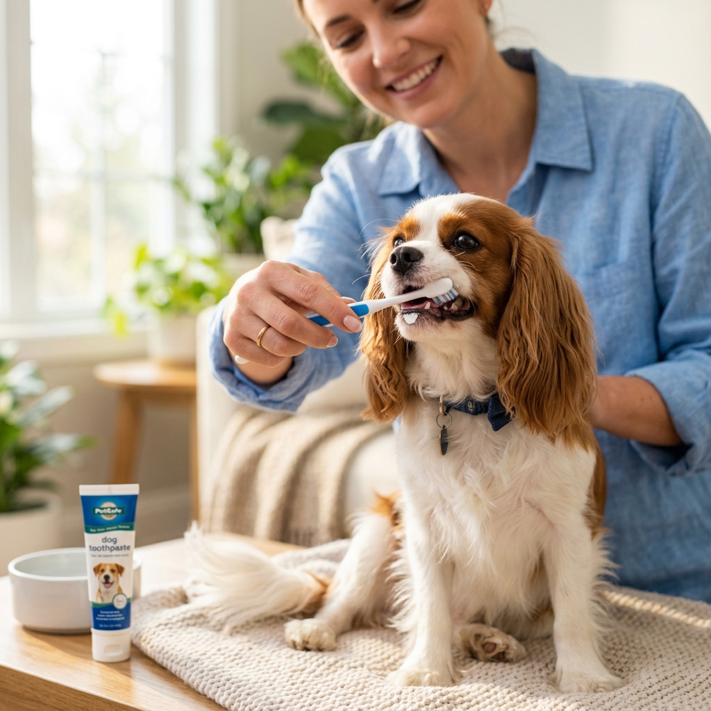
While annual professional dental cleanings under anesthesia remove calcified tartar, daily active home care represents the cornerstone of periodontal maintenance. Mechanical plaque removal via daily tooth brushing is the undisputed gold standard.
Pet-safe toothpastes must always be utilized; human toothpaste contains detergents and xylitol, a sugar substitute that causes profound hypoglycemic collapse and acute liver necrosis in canines. Veterinary toothpastes incorporate dual-enzyme systems (glucose oxidase and lactoperoxidase) that produce hypothiocyanite ions in the presence of salivary thiocyanate, naturally inhibiting oral bacterial proliferation.
Brushing technique should prioritize the buccal (outer) surfaces of maxillary premolars and molars at a forty-five-degree angle toward the gingival margin, as salivary duct openings deposit high concentrations of mineralizing salts adjacent to the upper fourth premolars. Introducing brushing gradually with positive reinforcement establishes lifelong cooperation without stress.
In addition to mechanical tooth brushing, veterinary antiseptic oral rinses containing 0.12 percent chlorhexidine gluconate offer powerful anti-plaque efficacy. Chlorhexidine exhibits strong substantivity—the unique biochemical ability to bind to oral hard and soft tissues and release sustained bactericidal concentrations over twenty-four hours. For pets suffering from early stage gingivitis, applying chlorhexidine rinses along the gingival sulcus significantly diminishes subgingival bacterial loads. Furthermore, professional dental barrier sealants, such as hydrophilic polymer formulations (SANOS) applied after ultrasonic scaling, form a durable barrier along the gingival margin, preventing plaque accumulation for up to six months.
5. Veterinary Oral Health Council (VOHC) Standards and Dietary Management
For pet parents struggling with daily tooth brushing, chemical and passive mechanical interventions provide meaningful adjunct protection. Caregivers should exclusively select products bearing the official seal of the Veterinary Oral Health Council (VOHC).
The VOHC is an independent scientific body that reviews rigorously controlled clinical trials to verify whether a product demonstrates a statistically significant reduction in plaque or calculus (typically a minimum of twenty percent reduction). VOHC-accepted categories include specialized dental kibble, edible rawhide chews, water additives, and topical oral gels.
Prescription dental diets function through unique physical kibble architecture. Unlike standard extruded kibble that shatters immediately upon contact, dental formulations feature enlarged kibbles with aligned fiber matrices that resist crumbling. The tooth penetrates deep into the kibble before it splits, mechanically squeegeeing plaque from the tooth crown. In addition, chemical calcium sequestering agents—such as sodium hexametaphosphate (HMP)—bind salivary calcium ions, preventing them from precipitating into hard calculus.
Disclaimer: The information provided is for educational purposes only. Always consult with a licensed veterinarian or board-certified veterinary dentist (DAVDC) for comprehensive oral evaluations and anesthetic dental procedures.